


 Foundational characteristics of cancer include proliferation, angiogenesis, migration, evasion of apoptosis, and cellular immortality. Find key markers for these cellular processes and antibodies to detect them.
 The SUMOplot™ Analysis Program predicts and scores sumoylation sites in your protein. SUMOylation is a post-translational modification involved in various cellular processes, such as nuclear-cytosolic transport, transcriptional regulation, apoptosis, protein stability, response to stress, and progression through the cell cycle.
 The Autophagy Receptor Motif Plotter predicts and scores autophagy receptor binding sites in your protein. Identifying proteins connected to this pathway is critical to understanding the role of autophagy in physiological as well as pathological processes such as development, differentiation, neurodegenerative diseases, stress, infection, and cancer.
> home > Products > Primary Antibodies > Antibody Collections > KD-Validated Antibodies > KD-Validated Anti-Arp3 Rabbit Monoclonal Antibody
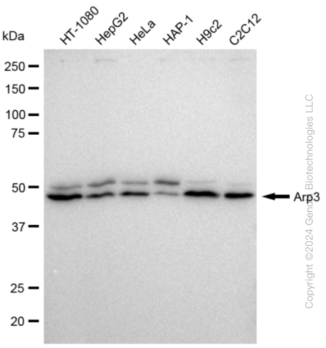
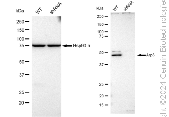
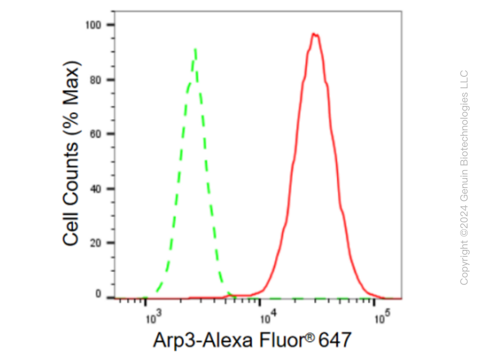
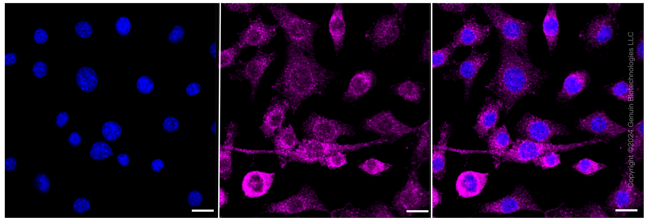
KD-Validated Anti-Arp3 Rabbit Monoclonal Antibody
Rabbit monoclonal antibody
- SPECIFICATION
- CITATIONS
- PROTOCOLS
- BACKGROUND

Application
| WB, FC, ICC |
|---|---|
| Primary Accession | P61158 |
| Reactivity | Rat, Human, Mouse |
| Clonality | Monoclonal |
| Isotype | Rabbit IgG |
| Clone Names | 23GB615 |
| Calculated MW | Predicted, 47 kDa; Observed, 47 kDa |
| Gene Name | ACTR3 |
| Aliases | ACTR3; Actin Related Protein 3; ARP3; ARP3 Actin Related Protein 3 Homolog; Actin-Related Protein 3; Actin-Like Protein 3; ARP3 (Actin-Related Protein 3, Yeast) Homolog; ARP3 Actin-Related Protein 3 Homolog (Yeast) |
| Immunogen | A synthesized peptide derived from human ACTR3 |
| Gene ID | 10096 |
|---|---|
| Other Names | Actin-related protein 3, Actin-like protein 3, ACTR3, ARP3 |
| Name | ACTR3 |
|---|---|
| Synonyms | ARP3 |
| Function | ATP-binding component of the Arp2/3 complex, a multiprotein complex that mediates actin polymerization upon stimulation by nucleation-promoting factor (NPF) (PubMed:9000076). The Arp2/3 complex mediates the formation of branched actin networks in the cytoplasm, providing the force for cell motility (PubMed:9000076). Seems to contact the pointed end of the daughter actin filament (PubMed:9000076). In podocytes, required for the formation of lamellipodia downstream of AVIL and PLCE1 regulation (PubMed:29058690). In addition to its role in the cytoplasmic cytoskeleton, the Arp2/3 complex also promotes actin polymerization in the nucleus, thereby regulating gene transcription and repair of damaged DNA (PubMed:17220302, PubMed:29925947). The Arp2/3 complex promotes homologous recombination (HR) repair in response to DNA damage by promoting nuclear actin polymerization, leading to drive motility of double-strand breaks (DSBs) (PubMed:29925947). Plays a role in ciliogenesis (PubMed:20393563). |
| Cellular Location | Cytoplasm, cytoskeleton. Cell projection. Nucleus. Note=In pre- apoptotic cells, colocalizes with MEFV in large specks (pyroptosomes) (PubMed:19109554) |
Research Areas
Citations (0)

Thousands of laboratories across the world have published research that depended on the performance of antibodies from Abcepta to advance their research. Check out links to articles that cite our products in major peer-reviewed journals, organized by research category.
Submit your citation using an Abcepta antibody to
info@abcepta.com, and receive a free "I Love Antibodies" mug.
info@abcepta.com, and receive a free "I Love Antibodies" mug.

Application Protocols
Provided below are standard protocols that you may find useful for product applications.
Abcepta welcomes feedback from its customers.
If you have used an Abcepta product and would like to share how it has performed, please click on the "Submit Review" button and provide the requested information. Our staff will examine and post your review and contact you if needed.
If you have any additional inquiries please email technical services at tech@abcepta.com.
$ 399.20

$ 149.00
Cat# AGI2347
Ordering Information
United States
AlbaniaAustraliaAustriaBelgiumBosnia & HerzegovinaBrazilBulgariaCanadaCentral AmericaChinaCroatiaCyprusCzech RepublicDenmarkEstoniaFinlandFranceGermanyGreeceHong KongHungaryIcelandIndiaIndonesiaIrelandIsraelItalyJapanLatviaLithuaniaLuxembourgMacedoniaMalaysiaMaltaMexicoNetherlandsNew ZealandNorwayPakistanPolandPortugalRomaniaSerbiaSingaporeSlovakiaSloveniaSouth AfricaSouth KoreaSpainSwedenSwitzerlandTaiwanTurkeyUnited KingdomUnited StatesVietnamWorldwideOthers
USA Headquarters
(888) 735-7227 / (858) 622-0099 or (858) 875-1900
Other Products
Shipping Information
Domestic orders (in stock items)
Shipped out the same day. Orders placed after 1 PM (PST) will ship out the next business day.
International orders
Contact your local distributors
